|
|||||||||

|
Projects: Embryo Development
|
||||||||||||||||

|
||||||||||||||||
| The Embryo Development is a comprehensive study of the growth process of the human embryo. We aim to completely label and visualize the developmental anatomy of the human embryo. | ||||||||||||||||
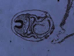
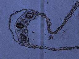
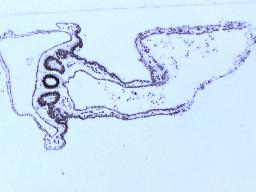
| The Visible
EmbryoTM project at the Human Developmental Anatomy Centre (HDAC)
offers a vast resource of microscopic sections of the human embryo
from Stage 1 - 23. Using select embryos from the Carnegie Collection
of 660 embryos, we aim to visualize the dynamics of tissue/organ growth.
This project is part of CieMed's vision of multi-scale
visulization and shape dynamics.
In addition to our collaboration with HDAC, a second research project in collaboration with the Dept. of Anatomy, National University of Singapore, and University of Alberta, Edmonton, Canada to quantify and visualize the growth of the central nervous system is also underway. News feature about our work on embryo development and anatomy visualization. |
||||||||||||||||
|
||||||||||||||||
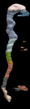
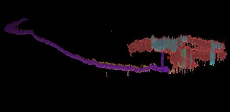
Stage-10
Embryo
Animated visualization of a late stage-10 Carnegie collection embryo. The skin, gut, and circulatory system fade away as the Spinal/Neural tube is exposed.
Animated visualization of a late stage-10 Carnegie collection embryo. The skin, gut, and circulatory system fade away as the Spinal/Neural tube is exposed.
Embryo:
Neural Tube
Animation of the Neural Tube of a Stage 10 Embryo. Highlighted parts are: Spinal cord, ventricles, and trigeminal ganglion.
Animation of the Neural Tube of a Stage 10 Embryo. Highlighted parts are: Spinal cord, ventricles, and trigeminal ganglion.
| Stage-10 Embryo | Embryo: Neural Tube | |||
| 4.3 MB mov | 609 KB mov |
| Profile | Research | Publications |